The reasons for organism’s aging
Effective anti-aging peptide Amatokin and Hexapeptide 20
Aging of a human organism is a normal biological function contributing to the progressive evolution of the species. Senility and the following death clean the population from ancestors and create the conditions for the descendants’ development – the carriers of new useful features. There are a lot of theories of ageing. The main idea of which is formed by two scientific theories. These are the theory of marginotomy and the theory of molecular, oxidative damage of cellular organelles by free radicals. The molecular, oxidative theory of aging. A lot of cells die premature because of a free radical attack (oxidant stress). The damaging effect of free radicals has been known for a long period of time. Free radicals injure cellular organelles causing the “breach” of a cell membrane, damage protein, amino acids, activate the peroxidation of lipids, and etc. Exactly free radicals are capable of damaging the tenderest cellular organelles – mitochondrions. As it is known, with advancing age the production of antioxidants reduces and the natural system of anti-oxidant protection becomes defective. The content of coenzyme Q, which is used to protect mitochondrions from antioxidants, decreases in mitochondrions because the production of energy, electrons’ transfer and formation of active forms of oxygen take place there. Of course, free radicals damage also cellular membranes, cellular organelles, DNA but the most important fact is that a mitochondrions’ damage switches on the programme of apoptosis or of natural cell death. As a result, a large quantity of cells which are viable and functional, have a big, unspent reserve of cells division and a high telomerase and which were far from aging, die. Together with them surrounding cells die as well because of apoptosis. The cells near a cell damaged by free radicals and which switched on the apoptosis programme start the apoptosis programme too because of “horror”. It brings, as it has already been mentioned above, to the cellular impoverishment of tissues, first of all of skin, and exactly this mechanism determines premature aging. The premature aging starts in an organism much earlier than it really should do. It is impossible to fight with the genetically determined aging but it is real to prevent fully the coming of the premature aging. The theory of marginotomy connects the aging with the contraction of the telomeric DNA. High differentiated, specialized cells of any organism’s tissue have a limited capability of proliferation. A gradual decrease of the cells’ proliferation rate causing its stop is called a cell or replicative aging. Aging cells progressively loose their function. Exactly they, staying in the tissues without division and regeneration, determine the aging elements display, because they are not replaced by young, normally functioning cells. They can’t proliferate because of the proliferation block which a telomere “switches on” having passed a necessary periodicity of cells division. A telomere is a measure of a cells’ proliferative potential. Chromosome ends are protected by the so-called tips – telomeres. With every cells division the end of a molecule as if breaks (by 50 – 200 nucleotides) because the apparatus of replication can’t produce a full-size copy of a DNA molecule, and also to avoid the chromosome fusion causing a genetic break. A telomere reduces with every cells division, and having reduced up to a certain length “switches on” the proliferation block and then the programme of apoptosis (natural cell death). Chromosomes end in the sequence ТТАGGG repeated in telomeres hundreds and thousands times. The loss of the end DNA makes impossible the endless proliferation. The chromosome shortening up to a certain size evokes the processes of cell aging. The number of replications passed by a cell up to the “switching on” of the proliferation block is different among different cells. Usually it is from 30 to 100 divisions. DNA is the only macromolecule having enough stability; it is the basis of such a mechanism. The cell which reduced its telomere up to the limit and switched on the proliferation block would not divide. It is necessary to reconstruct the telomere’s length for a cell not to loose its function and to continue dividing. This function is realized by the telomerase. The telomerase enzyme is not present in all cells of tissues and that is why it is impossible to make an aging cell proliferate. This enzyme is absent in differentiated, mature and specialized cells of organism’s tissues. An adult normally has telomerase only in gametes and precursor cells. To reconstruct the periodicity of cells division with a maximum short telomere of a cell is impossible because there is no telomerase in grown-up, differentiated, specialized cells of tissues. However, this process emerges in the organism much later than we really face with the displays of aging elements. Practically always an organism suffers from untimely aging. It happens because of the premature death of a large quantity of normally functioning cells that brings to the cellular impoverishment of tissues.The role of the immune system in the regulation of cells division
A humoral constituent of cell-cell interactions in the immune system is mediated by the products of interacting cells – by signal, informational, regulatory molecules – cytokines. They are albuminous and polypeptide products of activated cells of the immune system which are mediators of cell-cell communications (interactions) in the immune response, hemopoiesis and inflammation progress. They are effectors of some immune reactions and serve as a connecting-link between the immune and other systems of an organism. The value of cytokines goes beyond the scope of immunology because they play an important role in cells division, tissues’ regeneration, blood formation, and etc. Human skin is not only a protecting cover but also an active part of the immune system which possesses a feedback with it. It means that the state of skin integuments is regulated by the immune system and, vice versa, the immune system depends on functional activity of skin integuments. Quickly regenerating tissues such as blood and skin are in direct relation to the state of the immune system. Specialized skin cells interacting with each other participate in the formation of informational molecules and synthesis of cytokine cascades, and in the immune response. The immune system fulfills a lot of functions in an organism. The main ones are the fight against microbial agents, infectious agents, restoration of integrity and functioning of organism organs and tissues. The formation of a cytokine cascade, growth factors, informational molecules regenerating skin integuments and being in charge with reparative processes in skin and the whole organism depend exactly on the immune system. It is necessary to have growth factors and cytokines for the normal cells division. Many cytokines are not growth factors. Growth factors assist the DNA duplication in a cell and thereby bring it to the transition of the mitotic phase and to mitosis itself. Growth factors are necessary for cell life-support. For example, if cytokines and growth factors are removed from a cell culture, a cell not only will stop its division but will die. The immune system is responsible for normal cells division. That is why it is necessary for aging skin to support local immunity as a guarantee of normal cellular regeneration. Immune modulation (immune enhancement) of aging skin leads to the activation of cells division, to the normalization of cellular regeneration and to prophylaxis and treatment of skin age-related changes, to smoothing of aging development. The immunomodulating therapy started opportunely lets avoid premature aging and significantly delay genetically determined aging. Genetic and structural likeness of epidermis and thymus has been proved. Epidermal cells, like thymus, influence the maturation and differentiation of T-lymphocyte. It is the keratinocytes, which are generally considered as the keratin producers, are able to influence powerfully the biology of a T-cell. Skin keratinocytes influence the post-thymic stages of T-cells’ maturation. Keratinocytes, “a silent majority” of epidermis cells, are an important element of immunity. They not only provide the creation of keratinocyte blanket and hair coat on the body surface but also in certain conditions produce active substances being able to deeply affect the functioning of other skin cells. A certain “push” – the emergence of informational molecules signaling the necessity of cellular regeneration – is needed for this. Besides keratinocytes in the formation of the normal immune response Langerhan’s and Greenstein’s cells play an important role. For the last period of time a lot of informational molecules responsible for cell-cell interaction has been discovered. The peptide regulation which is one of prospective studies in the anti-age therapy has become widely spread. Such a therapy has a series of considerable advantages and, first of all, pathogenetic validity. With any aging (chronoaging or premature aging) the regulation disorder of informational molecules transfer between cells takes place that inevitably brings to the disorder of cells division. A lot of peptides realizing intercellular unions were defined from the tissues of a human organism. They are immunity inducers, peptides assisting cells division. There are also peptides responsible for vascular tone, mood, and etc. The contemporary science lets get such peptides not by means of extraction from tissues and organs now but by means of chemical, so-called peptide synthesis.Why is the peptide Amatokin important for aging skin?
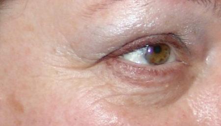
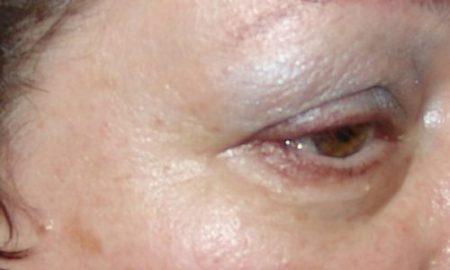
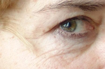
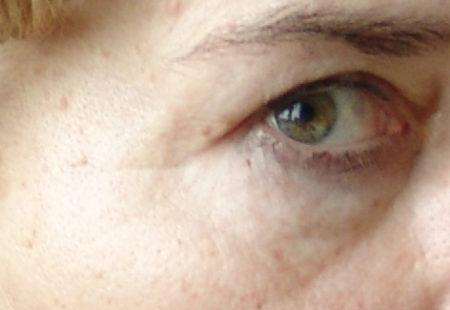
The peptide Amatokin restores normal cellular regeneration of skin. It does not care for only cellular regeneration but assists the normalization of cellular functions protecting a cell from death. It causes the emergence of new skin cells and takes care of aging ones. A lot of cells die prematurely because of a microcirculation disorder and free-radicals attacks. As a result of a microcirculation disorder the inflow of nutrients and oxygen to cells decreases that brings to accumulation of large quantities of acid metabolites and to production of free radicals. The peptide Amatokin will protect these cells from death and restore microcirculation. The peptide Amatokin influences the mitotic balance – the ratio of dividing, maturing, functioning and dying cells. Interacting with certain receptors the Amatokin regulates the most important functions: — restores a normal cells division cycle of mature, differentiated cells; — helps poorly differentiated precursor cells mature, turn into mature cells; — supports a high activity of nonspecific protective and reparation intracellular systems, increases cells’ survival rate and decreases the possibility of apoptosis (natural cell death) development of cells in the usual course of aging process. In the peptide Amatokin and Hexapeptide 20 make up everything what is necessary for aging skin for its restoration and regeneration, renewal normalization of a skin cellular structure, replenishment of the lack of young and active fibroblasts, cellular regeneration of keratinocytes. It is a powerful immunomodulating complex. The safety and efficiency of this complex has been proved. It does not have any side effects, does not develop an allergy, is not teratogenic, under no circumstance, even exceeding many times the recommended dosages, is nontoxic and harmless. The regulatory Amatokin peptide stimulates the proliferation and differentiation of stem cells, skin precursor cells and the formation of new, young skin cells (fibroblasts and keratinocytes). Also the Amatokin peptide takes care of aging cells. It restores the cascade of biologically active molecules which activate natural cytocinesis of both young and aging cells. It happens because of Amatokin’s influence on aging fibroblasts, keratinocytes, T-leucocytes, Langerhan’s cells and monocytes. When Amatokin interacts with certain receptors on outer membranes of aging cells, their activation starts. And aging cells restore their ability to induce a certain cascade of biologically active molecules causing the activation of precursors and other skin cells, and also of aging cells which have not used up their proliferation potential, normalize their cells division cycle. The unique Amatokin peptide is a signal molecule possessing an immunomodulating characteristic. With local application it renews a cellular strucutre, induces cells division, enhances the natural mechanism of reparation and regeneration, restores the cells division cycle in mature specialized cells, prevents cell death, and protects cells from damage. Amatokin allows to get new young fibroblasts and keratinocytes affecting those cells which are on a basal membrane, in the skin and hypodermic fatty tissue. Aging, as it has already been mentioned above, is a physiological process and skin does not give any signs about a disorder. It does not show distress signals, does not ask for help, does not say: “fix me” as it does a superficial wound. Loose skin and wrinkles’ emergence are normal a genetically programmed phenomenon. That is why it is necessary to help skin, to send it a signal molecule providing the launching of the whole complex of reparative processes. The creation of “a stress-like” situation and then the amplification of the natural process of skin reparation bringing to the renewal of skin cellular structure and a rejuvenating effect will happen. Amatokin launches a whole cascade of biological molecules being synthesized one after another at the place of its applying in the skin. It is a cascade of cytokines, growth rates and informational molecules. Precursor cells are highly sensitive to cytokines and growth factors. Mature differentiated cells of tissues are sensitive to growth factors only if they preserved the potential of divisions. Amatokin induces the synthesis of cytokines’ cascades such as IL-1, IL-3, IL-6, IL-8, and etc., which modulate a stress-like situation. The majority of them is antiphlogistic and enhances reparative processes. For example, the stem cell factor, GM-CSF, G-CSF, M-CSF induce a cascade of growth factors. The induction of the above-listed mediators takes place in the skin at the place of application, affecting the skin cellular elements (keratinocytes, fibroblasts, Langerhan’s cells, helper-inducer T cells, macrophages, monocytes). As a result the concentration of growth factors such as granulocytic, macrophage, granulocytic-macrophage, colony-stimulating ones increases in the skin. Amatokin displays activity only in the place of application and does not have a resorptive effect. With the Amatokin’s application we do not disturb anything determined by nature, only help activate a normal reparative function. IL-1 is one of the most important cytokines in our life which emerges with any tissue damage, with any intrusion of microorganisms and together with IL-2, IL-4, IL-6, IL-8 helps regenerate tissues, protects skin and activates reparative processes affecting skin cells, especially evident its effect on fibroblasts, Langerhan’s cells, T-lymphocytes (helper-inducer T-cells) that brings to immunity enhancement. Exactly IL-1 together with the tumor necrosis factor-alpha emerge the first in the response to the Amatokin application and assists to the synthesis of all the rest cytokines and growth factors in the skin, right in the place of application. IL-1 and the tumor necrosis factor-alpha activate the cascade of cytokines such as IL-3, IL-2, IL-4, IL-6, chemokine – IL-8 and the cascade of growth factors such as granulocytic-macrophage, granulocytic, macrophage colony-stimulating factors. IL-3 provides the support of stem and precursor cells proliferation. IL-3 determines the formation of mixed and immature colonies. IL-3 is mainly an emergency regulator showing its action in the stress or immune response. IL-3 is a growth factor for mast cells of mucous tunics (generating chondroitin phosphate) and enhances growth-stimulating activity of IL-4 with respect to mast cells of the connective tissue. If IL-3 is a growth factor for the youngest hematopoietic elements and stem cells, thus the granulocytic-macrophage colony-stimulating factor (GM-CSF), granulocytic colony-stimulating factor (G-CSF), macrophage colony-stimulating factors (M-CSF) support proliferation of more differentiated, though also young, precursor cells. Also GM-CSF affects the formation of specialized granulocytic and monocytic-macrophage colonies and the activity of mature monocytes of macrophages, granulocytes and eosinofils. Besides, this factor increases the macrophage’s generation of IL-1. GM-CSF also activates (helps synthesize) the stem cells factor. Like other hemopoietines, stem cells factor is produced first of all by stromal cells, especially by fibroblasts. Its main target cells are stem cells, early committed precursors of different types of cells. The stem cells factor affects stem cells and young representatives of hematopoietic type synergistically with IL-3, GM-CSF, G-CSF, M-CSF. They participate in supporting of proliferation of the youngest forms of precursors in skin, on a basal membrane, in hypodermic fatty tissue. Concerning mast cells it is the main growth factor and chemotactic agent. IL-8 or the neutrophil attraction factor (NAP-1) relates to the group of chemoattractive peptides – α-chemokines. IL-8 is produced by many types of cells and has evident anti-inflammatory features. The main biological effect of IL-8 is induction of chemotaxis of neutrophils, eosinophils, basophils and other cells of the immune system. IL-8 enhances neoangiogenesis in vivo and in vitro. Biological effects of IL-6 are similar to the effects of IL-1 and the tumor necrosis factor (TNFα). First of all, it is participation in realization of inflammatory and immune reactions and hematosis. Its effect on local manifestation of inflammation is similar to the IL-1 effect. Its characteristic is antiviral action. Being released a bit later than IL-1 and TNFα, IL-6 suppresses their generation (they, on the contrary, stimulate its generation), and that is way it relates to the cytokines finishing development of the inflammatory reaction. In the immune system the main target of IL-6 is B-lymphocytes. IL-6 is a cofactor of their proliferation and an independent differential factor. It evenly stimulates generation of immunoglobulin of all types. Besides, IL-6 is a growth factor of plasma cells and a hybrid. IL-6 affects precursor cells mainly as a cofactor assisting the effect display of IL-3, GM-CSF and M-CSF. As a result IL-6 enhances the formation of in vitro colonies of all types. Not possessing an independent radiation protective action, it increases this action in IL-1. IL-6 is an obligatory cytokine for the support of viability and proliferative potential of stem cells in a culture. So, IL-6 is a polyfunctional cytokine produced by fibroblasts, macrophages and other cells. According to its biological effect spectrum it is close to IL-1 and TNFα, takes part in inflammatory progress, immune reactions, hematosis regulation, serves as a growth factor for many types of cells, for fibroblasts and keratinocytes, participates in intersystem and cell-cell interactions. The content increase of these factors is a cascade of cytokines and growth factors necessary for effective cell regeneration of skin. The synthesis of such a cytokine cascade is a proof of Amatokin effectiveness. Exactly this factor brings to proliferation and differentiation of stem cells and precursors into mature, high-differentiated skin cells, restoring a necessary quantity of functioning cells. It is prophylaxis and fight against aging and age-specific cellular impoverishment. A cascade of these cytokines and growth factors indicates the Amatokin capability of influencing the proliferative potential of mature, differentiated cells, capability of restoring a cells division cycle, normalizing cell-cell interaction and preventing premature cell death. The content increase of growth factors and cytokines shows a strong immunostimulating effect of the peptide and also normalizes, rehabilitates a cells division cycle by means of regulating cell-cell interaction and supplying an outer cell membrane with powerful proliferative signals. Synthesized cytokines and growth factors interact with certain receptors on the outer surface of the cell membrane and reversibly change a receptor that assists the conducting of the proliferative signal to a cell and cells division. Granulocytic and macrophage colony-stimulating factors and also the granulocytic-macrophage colony-stimulating factor influence less differentiated precursor cells switching on proliferation and differentiation in them that causes a significant regeneration of skin cellular content.The study of microcirculation
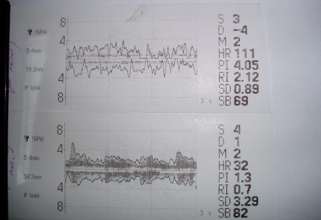
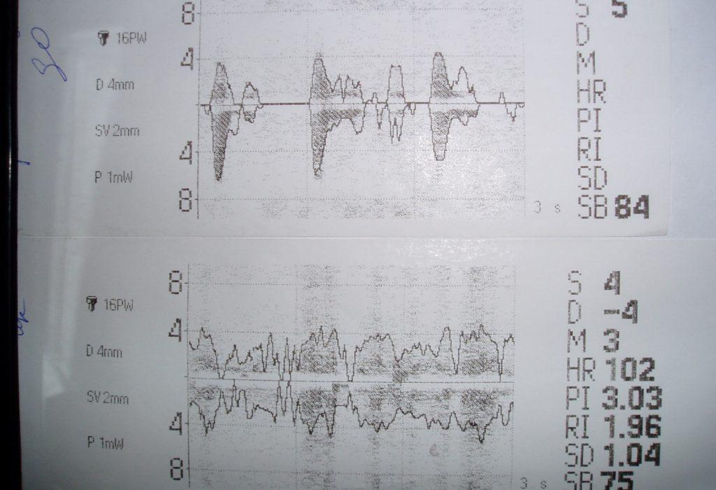
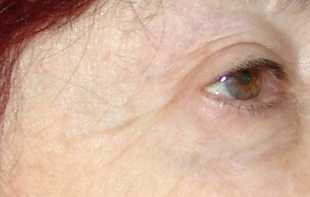
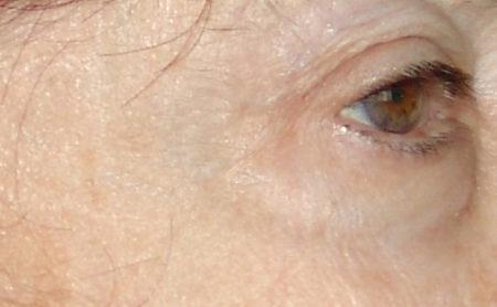
The Hexapeptide 20. Hexapeptide 20 lets eliminate a microcirculation disorder. Hexapeptide 20 protects microcapillaries from damage (vasoprotective activity), prevents from a microcirculation disorder and vascular permeability. Its main feature is the ability to restore the disordered microcirculation even in those microcapillaries where blood stasis has been observes for a long period of time. Hexapeptide 20 is the most effective stimulator of the blood flow capable of restoring it in the microcirculation after the influence of a damaging factor (ultraviolet radiation, toxins, malnutrition, and etc.). The recovery of blood flow takes place through the regulation of vascular tone. There where capillaries were narrowed and closed they open and the microcirculation is restored after the usage of Hexapeptide 20. There where capillaries were widened they narrow up to the normal size of lumen after the peptide’s application that brings to the recovery of microcirculation and eliminates blood stasis, paralytic vasodilatation of microvessels disappears, normal blood circulation restores in these vessels of microcirculation. The peptide rehabilitates not only blood inflow in arterioles but also reconstructs blood outflow in venules and regulates their lumen. That is microcirculation restoration. After the Hexapeptide’s 20 application on the skin the following happens: — the increase of blood circulation in microcapillaries, — the increase of functioning microcapillaries, — the restoration of blood flow in the microcapillaries being earlier in stasis. An ultrasound Doppler device of the continuous-pulse analyzer-monitor of intracranial and peripheral circulation “Angiodin-PC” was used. In all there were 25 studies. The researches were carried out on women at the age of 35-55 years. The state of microcirculation before the Hexapeptide 20 application and in 20 minutes after its usage was measured. The following results were received: Blood circulation increased by 1,8 times among 85% of women and by 1,2-1,4 times among all the rest. At the same time normalization of the microcapillaries diameter takes place what is fixed in the edge flatness of the curve (top and bottom), and also the increase of blood supply of the researched part is observed that is expressed in the intensification of graph coloration. Before: After 10 minutes: After 20 minutes:At the top – before the Hexapeptide 20 application, At the bottom – after the Hexapeptide 20 application.
